Following the Fluorescent Clues of Microplastics
Mansfield
Posted
Microplastics are notoriously hard to find, but a student–faculty team at Commonwealth University–Mansfield has been busy building tools to track them in living tissue to better understand their health risks.
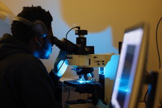
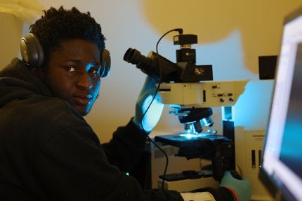
MANSFIELD — Under the cool blue glow of a fluorescent microscope, Jay Diya '25 scrolls through images of mouse tissue, circling bright flecks that don’t belong there — microplastics that a Commonwealth University-Mansfield research team is determined to track, count, and understand.
Microplastics, fragments smaller than a grain of sand, have become one of the most urgent and elusive subjects in modern environmental and biological research. They are everywhere … in water, soil, food, and increasingly, within living organisms.
But understanding where microplastics go and what they do once they get there requires more than curiosity. Diya, a biology graduate student, has been up to the challenge.
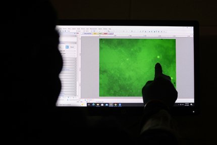
“I’m using enzymes to digest mammalian tissue without harming the microplastics, so we can isolate and quantify them,” Diya explains, standing beside a fluorescent microscope that casts an otherworldly glow across prepared samples.
His work focuses on a deceptively complex question of how you isolate microplastics from mammalian tissue without destroying them in the process. The answer, it turns out, is not straightforward.
“Most reagents that digest tissue also digest plastic,” says Elaine Farkas, associate professor of chemistry and physics and one of the project’s driving forces. “That’s really the chemistry problem.”
The challenge of understanding microplastic behavior in living systems has shaped Mansfield’s research trajectory since 2018, when standout undergraduates Katherine Thompson ’20 and Cody McUmber ’21 helped launch the initiative as cell and molecular lab assistants. Their early contributions established the groundwork for what has become one of the campus’s most ambitious multidisciplinary projects.
McUmber played a particularly pivotal role in the project’s formative years by conducting the initial mouse study, gavage‑feeding microplastics to assess physiological responses, according to Kristen Long, associate professor of biological sciences and key faculty collaborator and research adviser.
McUmber was also the first to expose bone marrow–derived cells to microplastics and the first to examine liver histology for immune‑cell changes. His work opened the door to deeper immunological questions the team continues to explore.
As the project expanded, so did the roster of contributors.
Kade Showers ’24/M25, a recent biology graduate student, built the experimental foundation now being refined and optimized by current researchers. While pursuing graduate studies at the Medical University of South Carolina, according to Long, he continued to support the project by running flow cytometry for Showers’ samples, ensuring continuity and precision in the data pipeline.
Marvens Ravix ’25 added another critical layer by quantifying and identifying immune cells within liver tissue, helping the team map how microplastics interact with and potentially disrupt immune pathways.
What began as a timely, student‑driven inquiry has evolved into a collaborative effort bridging biology, chemistry, and physics — a rare and ambitious undertaking for a campus laboratory. At its core, the research centers on two fundamental questions of where microplastics accumulate in living systems and what happens once they arrive.
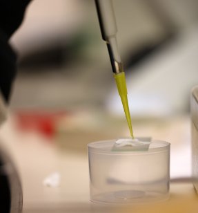
To answer those questions, Mansfield researchers must first find and measure the plastics inside biological tissue. “It’s like looking for a needle in a haystack,” Farkas says.
Instead of using harsh chemicals that can damage the plastics, Diya is trying gentler enzyme methods to break down animal tissue while keeping the microplastics intact.
“The goal is to find the best enzymatic digestion protocol,” Diya says. “Something that works across mammalian tissues and is easy to replicate.”
Once the tissue is digested, the next step begins.
Under a fluorescent microscope, Diya analyzes the remaining material, searching for tiny particles that emit a distinct glow. Each image becomes data. Each data point contributes to a larger picture. One that could reveal how microplastics move through organs, where they accumulate, and what biological effects they may trigger.
“If we can quantify them,” Diya says. “… we can start to see where they tend to bioaccumulate and what effects they have on those organs.”
It’s meticulous work that requires hours of imaging, cataloging, and analysis. But for Diya, the process is as valuable as the outcome. He began his research journey early, assisting on multiple projects as a freshman before launching his own work as an upperclassman.
That early exposure, according to Diya, fundamentally changed how he approaches problems.
“It gives you a brand-new way of thinking,” Diya says. “You start learning analysis before you even take the class. You understand how to form hypotheses, test them, and interpret results.”
That mindset now extends beyond the lab. Diya plans to attend physician assistant school once he completes his master’s degree program to further pursue his interests in hospital medicine and dermatology.
While his future may not center on microplastics, the skills he’s developing — data interpretation, image analysis, scientific reasoning — will carry forward.
“If I need to look at medical images or analyze cells, I’m already comfortable with that,” Diya says. “This experience builds that foundation.”
Farkas says the transformation in students — at both the graduate and undergraduate level — is one of the most important outcomes of the project.
“The techniques are useful, but it’s also about confidence,” Farkas says, “Students realize they can walk into a lab, think independently, and solve problems. They’re not just following a template. They’re doing real science.”
That sense of ownership is amplified by the project’s originality.
“There’s still so much we don’t know,” Farkas says. “Environmental studies tell us where plastics are, but controlled dose studies help us understand what they’re doing inside organisms.”
Over the past eight years, the Mansfield team has explored multiple models, from plants to invertebrates to mammals, reflecting the broad scope of the issue. Microplastics move across ecosystems, food chains, and biological boundaries. And measuring them remains one of the field’s greatest challenges.
“We’re trying to refine the chemistry enough that we can use flow cytometry to get accurate counts,” Farkas says. “That’s something that hasn’t really been done yet.”
Because ultimately, this research is not just about microplastics. It’s about understanding how human-made materials interact with living systems and what that means for health, environment, and future policy.
Since early spring, Diya has continued scanning his samples, each fluorescent signal a small piece of a much larger puzzle. The work is slow. The questions are complex. The answers are not guaranteed.
“You’re not just repeating something,” Farkas says. “You’re figuring it out.”
And for students like Diya, that’s where science truly begins.